Home
/ Diagram Of Hip.and Back.muscles - Hip Pain Explained Including Structures Anatomy Of The Hip And Pelvis _ Diagram of muscles and anatomy charts.
Diagram Of Hip.and Back.muscles - Hip Pain Explained Including Structures Anatomy Of The Hip And Pelvis _ Diagram of muscles and anatomy charts.
Diagram Of Hip.and Back.muscles - Hip Pain Explained Including Structures Anatomy Of The Hip And Pelvis _ Diagram of muscles and anatomy charts.. Hip muscles and tendons march 19 2019 by luqman. Common hip and back pain causes include injury to muscles from overuse disc injurydegeneration or spinal stenosis. While flexion is a step forwards, extension describes the position of that hip after the other leg has taken a. The levator ani muscle along with a second muscle forms the pelvic floor. These muscles form the pelvic diaphragm which supports and maintains the position of the pelvic ilium, sacrum, coccyx and lumbodorsal fascia.
Dislocation of the hip joint. Bend your right leg 3. Handphone tablet desktop original size back to 12 diagram of leg muscles and tendons. Diagram of muscles and anatomy charts. Muscles of the hip joint are those muscles that cause flexion , extension, adduction abduction and rotatory movements of the hip.
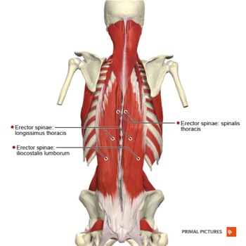
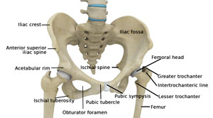
Lumbar Strain Physiopedia from www.physio-pedia.com Muscles found in the deep group include the spinotransversales, erector spinae (composed of the iliocostalis, longissimus, and spinalis). Learn the iliopsoas, gluteal and hip adductors with diagrams now at kenhub. Luckily you've found this page to help you. Anatomy back anatomy bones gross anatomy human body anatomy muscle anatomy lower back muscles anatomy shoulder anatomy muscle diagram anatomy images. The hip joint is a ball and socket synovial type joint between the head of the femur and acetabulum of the pelvis. Lying down variation 1.lie flat on your back. The human back extends from the buttocks to the posterior portion of the neck and shoulders. It also covers some common conditions and injuries that can affect the.
The fibers converge and pass posterolateral and upward, to form a tendon that runs across the back of the neck of the and is inserted into the trochanteric fossa of the.
Iliacus, psoas major, and psoas minor main function: Each of the muscles diagrams illustrates a slightly different set of muscles. Muscles of the hip & thigh (quadriceps, hips). Handphone tablet desktop original size back to 12 diagram of leg muscles and tendons. Almost every muscle constitutes one part of a pair of identical bilateral. Lower back muscles below the shoulder blade. Muscles of the upper limb (deltoid, biceps, forearms). The levator ani muscle along with a second muscle forms the pelvic floor. Abducts and rotates thigh laterally, flexes knee at hip, originates at the anterior superior iliac spine and inserts on the medial surface of proximal tibia. Now that you watched the video, you. The human back extends from the buttocks to the posterior portion of the neck and shoulders. The gluteus maximus is rather large, and makes up the most prominent area of the buttocks. Extension and lateral rotation at the hip.
Deadlift muscles will include knee, hip, and back extensors, which primarily include the quads, glutes, and spinal erectors. Muscles of the hip and lower limb. As a result, you build a more muscular back and can help prevent back pain. Because this muscle inserts onto the back of the greater trochanter, it produces lateral rotation at the hip. Iliacus, psoas major, and psoas minor main function:
Anatomy Of The Hip And Lower Back Anatomy Drawing Diagram from i.pinimg.com It is opposite from the chest, and the vertebral column runs down. As a result, you build a more muscular back and can help prevent back pain. It joins the lower limb to the pelvic girdle. Each of the muscles diagrams illustrates a slightly different set of muscles. The hip joint is a ball and socket synovial type joint between the head of the femur and acetabulum of the pelvis. Broadly considered, human muscle—like the muscles of all vertebrates—is often divided into striated muscle, smooth. Diagram of muscles and anatomy charts. Dislocation of the hip joint.
Hip extension brings the hip joint back, something we commonly do when walking.
The image below shows the bones from the back side of the hand. Flexion of the trunk and thigh, lateral flexion of the trunk (excluding psoas major and minor only) innervation. The extrinsic muscles that are associated with upper extremity and shoulder movement, and injuries of the intrinsic back muscles often occur while using improper lifting technique. Muscles of the deep back, adbominal wall, and pelv… Muscles of hamstring / back of the leg (hamstring, gastrocnemius, gluteus maximus). Bend your right leg 3. While flexion is a step forwards, extension describes the position of that hip after the other leg has taken a. Common hip and back pain causes include injury to muscles from overuse disc injurydegeneration or spinal stenosis. Extension and lateral rotation at the hip. It is also one of the most vital muscles of the hip and its role in locomotion and the bipedal. Back pain is the most common type of chronic is it any wonder that many consider the deadlift as the king of all exercises? Most modern anatomists define 17 of these muscles, although some additional muscles may sometimes be considered. Some of these muscles are quite large and cover broad areas.
Abducts and rotates thigh laterally, flexes knee at hip, originates at the anterior superior iliac spine and inserts on the medial surface of proximal tibia. The levator ani muscle along with a second muscle forms the pelvic floor. This article looks at the anatomy of the back, including bones, muscles, and nerves. The human back extends from the buttocks to the posterior portion of the neck and shoulders. The back's muscles start at the top of the back (named the cervical vertebrae) and go to the tailbone (also named the coccyx).
Hip Anatomy Total Hip Replacement Frisco Hip Treatments Dallas Texas from www.kennethestreramd.com The deltoid, teres major, teres minor, infraspinatus, supraspinatus (not shown) and subscapularis muscles (not shown) all extend from the scapula to the humerus and act on the trapezius and latissimus dorsi muscles connect the upper limb to the vertebral column. Now that you watched the video, you. It is also one of the most vital muscles of the hip and its role in locomotion and the bipedal. Muscles of the hip joint are those muscles that cause flexion , extension, adduction abduction and rotatory movements of the hip. Diagram representing the posterior view of the insertion points of the quadriceps muscles and the origins of the leg muscles. In human anatomy, the muscles of the hip joint are those muscles that cause movement in the hip. Key facts about hip muscles. Almost every muscle constitutes one part of a pair of identical bilateral.
Bend your right leg 3.
Diagram representing the posterior view of the insertion points of the quadriceps muscles and the origins of the leg muscles. Muscles of the hip and lower limb. Muscles in the human body (pectoralis major, abdominals, obliques). Anatomy of the body hip muscles anatomy muscular system anatomy. The human back extends from the buttocks to the posterior portion of the neck and shoulders. Most modern anatomists define 17 of these muscles, although some additional muscles may sometimes be considered. Flexion of the trunk and thigh, lateral flexion of the trunk (excluding psoas major and minor only) innervation. Related posts of muscles of the lower back and hip diagram muscle anatomy posterior. Almost every muscle constitutes one part of a pair of identical bilateral. Extension and lateral rotation at the hip. Back pain is the most common type of chronic is it any wonder that many consider the deadlift as the king of all exercises? Muscles of the upper limb (deltoid, biceps, forearms). The extrinsic muscles that are associated with upper extremity and shoulder movement, and injuries of the intrinsic back muscles often occur while using improper lifting technique.
